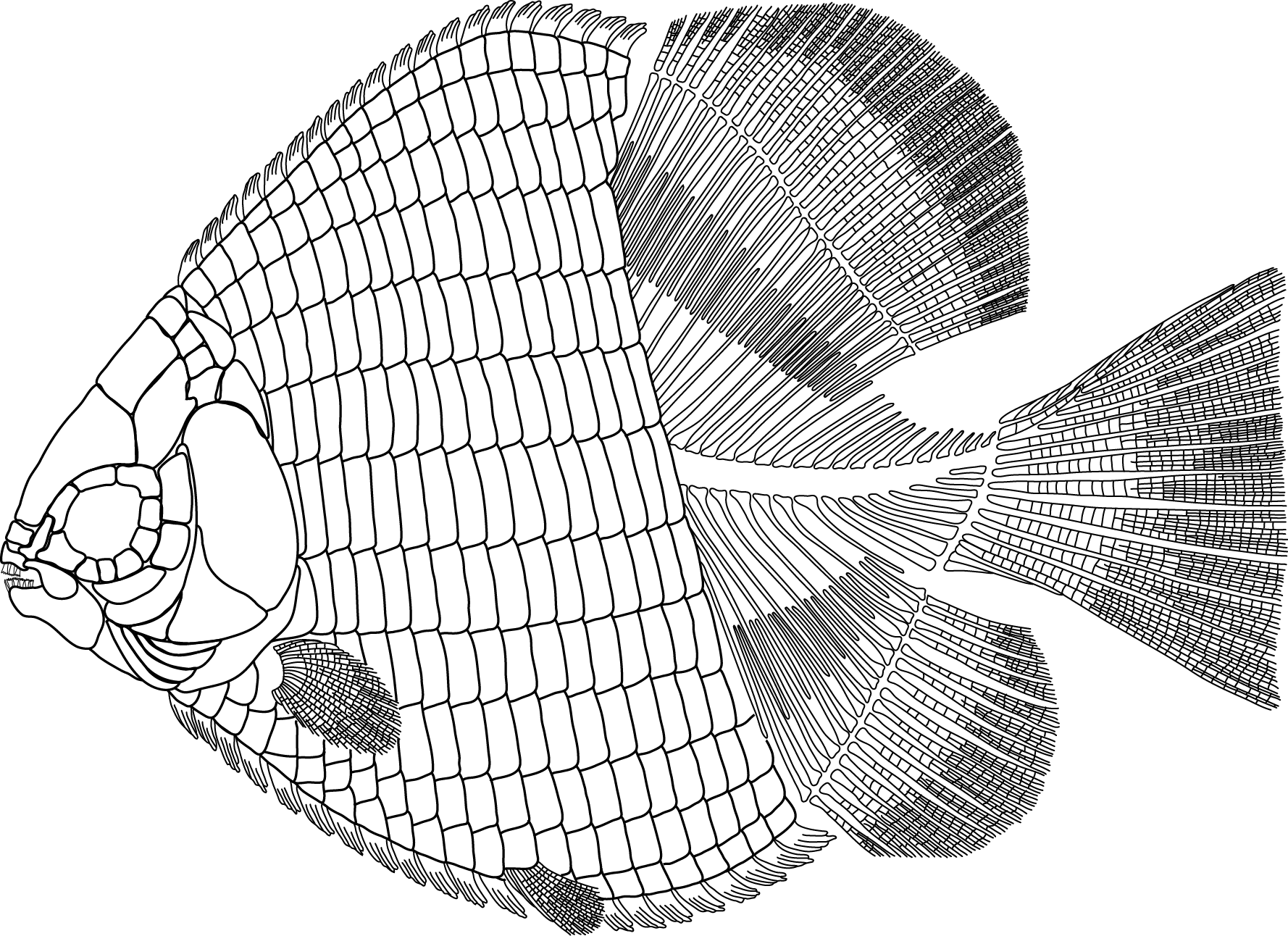
The (Now Older) Age of Fishes: New bony fish from the Silurian of China | PLOS Paleo Community

This is a blog post I wrote for the PLOS Paleo Community, and was published in March 2017. I am archiving it here on my personal website. The text has been slightly modified. You can see the original post here.

For all of the love and popularity that the “Age of Dinosaurs” receives, we wouldn’t be where we are without evolutionary innovations that occurred during the “Age of Fishes.” The Devonian Period (419.2–358.9 million years ago) witnessed a great increase in global abundance and diversity of jawed vertebrates. But a recent study presents evidence that early jawed fishes may have evolved earlier, in the Silurian Period (443.7–419.2 million years ago), with China at its epicenter.

The study, published in PLOS ONE by Brian Choo from Flinders University, Australia, with colleagues from the Institute of Vertebrate Paleontology and Paleoanthropology, China, describes a new fossil from the Kuanti Formation of Yunnan, southwestern China. The fish, dubbed Sparalepis tingi, is based on a beautifully-preserved and articulated partial specimen, and supports the idea that this area of Asia may represent an early boom of diversification of bony fishes, long before the Devonian “Age of Fishes.”

The holotype of Sparalepis ting (V 17915), from Choo et al. 2017.

The Kuanti Formation (~423 Ma) is rich in marine fossils: corals, molluscs, trilobites, as well as several various groups of fishes, including placoderms, acanthodians, and osteichthyans. Only until recently, however, the knowledge of fishes from this formation was based on highly fragmentary material. For example, one bony fish from the Kuanti, Naxilepis gracilis, is based solely on isolated scales. Another osteichthyan, Megamastax amblyodus, is large but fragmentary.

Then in 2009, Min Zhu (second author in this study) and colleagues described Guiyu oneiros, the oldest articulated bony fish with a mosaic of gnathostome characteristics, including some characteristics that were once attributed solely to placoderms. In 2013, Zhu et al. then published Entelognathus, a maxillate placoderm. These taxonomic studies are based on fantastically-preserved specimens from the Kuanti Formation, and with this study (published in 2017), Sparalepis becomes the third articulated specimen from the Kuanti Formation, and only second known articulated Silurian osteichthyan.

Diagrammatic reconstruction of Sparalepis, and examples of scales preserved. From Choo et al. 2017

Sparalepis is named after the Sparabara infantry during the Persian Empire due to the similarity of the shape of the scales of the fish to the shape of the wicker sheilds carried by the Sparabara. The scales are particularly tall, thick, and narrow with distinct interlocking articulation mechanisms. Sparalepis is represented by a partial postcranium, body scales, and some fin elements. The pectoral girdles bear large fin spines, as well as large dorsal fin spines beautifully preserved in the holotype.

A lot of these characters observed in Sparalepis are shared with Guiyu, in particular the spine-bearing pectoral girdle and placoderm-like, dermal pelvic girdle. These articulated specimens from the Silurian helped the researchers solve another fishy mystery, by helping them definitively identify isolated elements from the Devonian Xitun Formation in Yunnan, attributing them to the enigmantic taxon Psarolepis. (Zhu et al. 2012)

So what sets Sparalepis aside from Guiyu and Psarolepis as a distinct species? Sparalepis possesses prominent linear ridges and pore openings on the dermal surfaces of all of the larger bones and median scutes are unique to Sparalepis and lacking in Guiyu. The scale ornament of Sparalepis and Guiyu are similar to each other, and different of that seen in Psarolepis.

What is important to note about these fishes is that they display characteristics that were once thought to be isolated to placoderms. The mosaic of characters seen in Sparalepis and Guiyu has caused questions regarding previous evolutionary hypotheses of relationships between placoderms, sarcopterygians, and actinopterygians.

The team sought to address the relationships of these enigmatic fishes, and provided a dataset to test their phylogenetic hypothesis. Their results suggest that Sparalepis is closely related to Guiyu, Achoania, and Psarolepis; this group is recovered at the base of Sarcopterygii, in a clade that this study is putatively calling “psarolepids.” This study also provides additional support to the hypothesis that the ancestral species to gnathostomes likely possessed a placoderm-like bodyplan, and that characteristics seen in stem sarcopterygians like Sparalepis are likely plesiomorphic conditions.

So this study is helping to narrow a wide morphological gap in the early history of jawed vertebrates. And with the evidence provided by this study, we now know that that history began earlier than we thought. Thanks to Sparalepis and Guiyu, we can see that the “Age of Fishes” arrived early during the SIlurian in China.

References:

Choo B, Zhu M, Qu Q, Yu X, Jia L, Zhao W (2017) A new osteichthyan from the late Silurian of Yunnan, China. PLoS ONE 12(3): e0170929. doi:10.1371/journal.pone.0170929 

Zhu M, Zhao WJ, Jia LT, Lu J, Qiao T, Qu QM (2009) The oldest articulated osteichthyan reveals mosaic gnathostome characters. Nature 458: 469–474. doi: 10.1038/nature07855

Zhu M, Yu XB, Ahberg PE, Choo B, Lu J, Qiao T, et al. (2013) A Silurian placoderm with osteichthyan- like marginal jaw bones. Nature 502: 188–193. doi: 10.1038/nature12617 PMID: 24067611 

Zhu M, Yu XB, Choo B, Qu QM, Jia LT, Zhao WJ, et al. (2012) Fossil fishes from China provide first evi- dence of dermal pelvic girdles in osteichthyans. PLoS ONE 7: e35103. doi: 10.1371/journal.pone. 0035103 PMID: 22509388 

via The (Now Older) Age of Fishes: New bony fish from the Silurian of China | PLOS Paleo Community

Alamosaurus: how this massive titan’s neck is impacting relationships of titanosaurs | PLOS Paleo Community

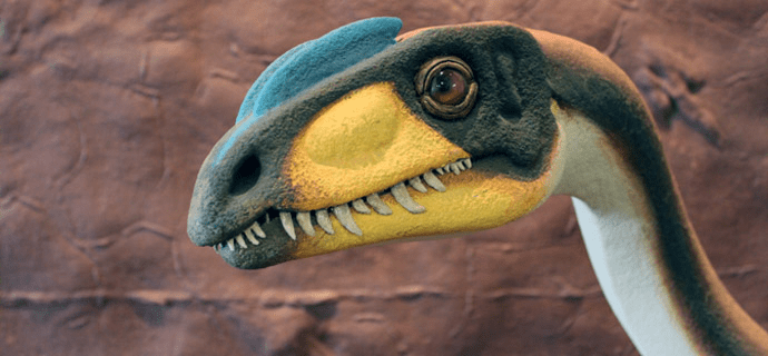
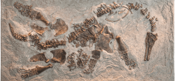
This is a blog post I originally wrote for the PLOS Paleontology Community blog, and am archiving it here on my personal website. You can find the original post here.

Remember the Alamo? Well, it’s easy to forget when you are staring at this massive dinosaur. It makes that Tyrannosaurus look like a puppy in comparison.

21584893173_a286943531_o
Alamosaurus on display at the Perot Museum. Photo by Sarah Gibson, taken during the SVP opening reception, October 2015.

At least that was how I felt when seeing the Alamosaurus on display at the Perot Museum of Nature and Science. I, along with hundreds of paleontologists at the 2015 Society of Vertebrate Paleontology meeting in Dallas, got to admire this beautiful behemoth at the opening social. Alamosaurus has a great and complex history to match its size, and now, with a new study by Ronald S. Tykoski and Anthony R. Fiorillo published open access this week in the Journal of Systematic Paleontology, Alamosaurus now comes with a more complete anatomical and morphological description, and a new hypothesis of how Alamosaurus relates to other titanosaurs.

“Giant sauropods like Alamosaurus have amazed people since the 1800s.  Their sheer size boggles the mind, and they have forced scientists to re-think the physical limits of land-living animals,” said Tykoski.  “The fossils described in our paper reveal new details about the last sauropods in North America, which helps us better understand who Alamosaurus was related to and how this species made it to southern North America by 67 to 66 million years ago – just in time to go extinct at the end of the Cretaceous!”

econtent.unm.edu
The Ojo Alamo Trading post circa 1911, before Alamosaurus was even discovered. Bones be out there, somewhere… Image courtesy the Farmington Museum

Now, Alamosaurus was not named after the THAT Alamo. Rather it was named for the Ojo Alamo trading post in New Mexico, where bones of the sauropod were first discovered in the 1920s. Since then, other remains have been discovered in Utah, Texas, and New Mexico. Most of these discoveries, however, were incomplete sections of the dinosaur, and so the whole picture remained elusive and its relationship to other titanosaurs was difficult to interpret.

In 1997, a joint team of paleontologists from the University of Texas at Dallas (UT-D) and the Perot Museum of Nature and Science found additional remains of Alamosaurus in Big Bend National Park. The scientists and volunteers were excavating a site that produced parts of several immature sauropods when Dana Biasatti, then a student at UT-D, came upon the remains of an adult titanosaur a few hundred yards away. The team was stunned. The nine cervical (neck) vertebrae were the first articulated series of adult Alamosaurus neck bones ever found. The fossils of Alamosaurus from Big Bend National Park currently represent the biggest dinosaurs discovered in Texas.

It would be another four years before they were able to excavate the remains, however. Working with the National Park Service and a helicopter service, several blocks, some weighing over a ton, were airlifted out to a flatbed truck about a mile away, and then transported over 500 miles to the Perot Museum in Dallas. The bones have been prepared and are now on display under the reconstructed Alamosaurus in the museum, which spans over 25 feet.

Figure-5.-Jacket-removal-from-Big-Bend-768x1156
Airlifting the remains of Alamosaurus out of Big Bend National Park. Image courtesy the Perot Museum of Nature and Science.

“This remarkable discovery illustrates the importance of America’s public lands as places where scientists have access to perform research that benefits everyone,” said Cindy Ott-Jones, Superintendent of Big Bend National Park. “While Big Bend National Park is a place that many people enjoy for its scenery and recreational opportunities, visitors should know that a tremendous amount of scientific research is also performed in the park.”

Because of the new anatomical and morphological information provided in the study, the authors were able to propose a a more robust hypothesis of evolutionary relationships of Alamosaurus to other sauropod dinosaurs. And the results of the phylogenetic analysis were novel; the researchers recovered Alamosaurus to the Lognkosauria clade, which includes the genera Futalognkosaurusand Mendozasaurus, both massive titanosaurs from the Cretaceous of Argentina. This new proposed relationship is supported by three shared derived characters—all characters based on the morphology of the cervical vertebrae, made possible by this discovery.

This study also has paleobiogeographical implications, mainly in why Alamosaurus is where it is at the time it is. Tykoski and Fiorillo (2016) discuss several scenarios depending on different studies, and in one scenario suggest a northward dispersal by the ancestors of Alamosaurus from South America—a hypothesis which matches their phylogeny, as its closest relatives proposed by this study are found in Argentina, it is likely their common ancestor was also from South America.

This paper is significant, and has been years in the making. The new data provided by Tykoski and Fiorillo (2016) illuminates possible evolutionary relationships of titanosaurs, and gives Alamosaurus a place on the family tree.

Reference:

Ronald S. Tykoski & Anthony R. Fiorillo (2016): An articulated cervical series of Alamosaurus sanjuanensis Gilmore, 1922 (Dinosauria, Sauropoda) from Texas: new perspective on the relationships of North America’s last giant sauropod, Journal of Systematic Palaeontology, DOI: 10.1080/14772019.2016.1183150

This post was modified from materials provided by the Perot Museum of Natural History and Science

via Alamosaurus: how this massive titan’s neck is impacting relationships of titanosaurs | PLOS Paleo Community

Track Makers in Southern Utah: The St. George Dinosaur Discovery Site | PLOS Paleo Community

Featured image: Megapnosaurus model on display in front of Grallator-type dinosaur tracks at the St. George Dinosaur Discovery Site at Johnson Farm. Photograph by Sarah Gibson.

This is a post that I originally wrote for the PLOS Paleontology Community blog in May of 2016. I am archiving it here on my website, so some of my references to the 2016 SVP meeting in Salt Lake City are a bit outdated. Nevertheless, you’ll get the gist about this awesome site. You can find the original blog post here.

UPDATE August 22, 2016: It is with great sadness that we announce the passing of Dr. Sheldon Johnson last week from leukemia. You may find his obituary here, and please consider donating to the DinosaurAh!Torium Foundation, which helps fund research and preservation of tracks at the St. George Dinosaur Discovery Site at Johnson Farm.

When it comes to absolutely amazing paleontological resources, Utah arguably reigns supreme within the United States (I may be a bit biased). And with the upcoming Society of Vertebrate Paleontology meeting taking place in Salt Lake City, paleontologists and paleo enthusiasts will be flocking to the Beehive State to discuss and share the latest breakthroughs in the field. To those coming to the meeting this October, one thing I cannot stress enough: do not miss what Utah has to offer in terms of spectacular fossil sites and museums. It will be difficult to avoid, as the SVP host committee this year is offering up eleven (eleven!!!) field trips to different parts of the state to see everything from the Triassic-Jurassic transition to Mesozoic dinosaurs to Eocene fishes to Pleistocene shorelines, and more.

If you’d rather go rogue and visit some places on your own, you are greeted with a plethora of options: at least a dozen museums, five national parks and many more national monuments, and some beautiful in situ fossil localities open to visitors. I recently visited one of the more recent additions of paleo places to visit in Utah, and it’s one of my favorites as well because, well, I worked there for two years! Let me make the case for why you should consider a jaunt down to St. George, Utah and view some of the most spectacularly preserved dinosaur tracks in North America.

sheldon
Dr. Sheldon Johnson pictured with one of the dinosaur track blocks in 2000. Image courtesy Andrew R. C. Milner.

Located at the southwestern tip of the state, the St. George Dinosaur Discovery Site at Johnson Farm(hereafter called SGDS) was only very recently discovered in 2000, when landowner and retired optometrist Sheldon Johnson was clearing some land for development. Overturning some large sandstone blocks, he observed natural casts of dinosaur footprints that were so detailed, he initially thought he had an actual dinosaur preserved in the rock with its foot hanging out. As he moved and overturned more blocks, he quickly realized he was looking at exquisite dinosaur trackways.

Now, here is where Dr. Johnson did a wonderful thing. Rather than destruction of the tracks in order to develop the land, he called local geologists and paleontologists, who quickly came and assessed the site. They found more of the tracksite in situ, and realized that they were dealing with multiple layers of dinosaur tracks, as well as invertebrate traces, body fossils, stromatolites, plants, and more. The City of St. George hired Andrew R.C. Milner to document and preserve the site, along with help from a dedicated group of volunteers, the Utah Friends of Paleontology, over the next few years. And working with the City of St. George, the Johnsons donated the land to preserve the site, and a museum was built over the bulk of the tracksite in 2005, still in its original location on the Johnson property.

Sixteen years since the initial discovery, the SGDS is operating better than ever, with over 38,000 visitors per year, according to Liz Freedman-Fowler, the Executive Director of the SGDS. The museum is now autonomous and is managed by the non-profit Dinosaur Ah!Torium Foundation. Andrew R.C. Milner continues to research, curate, and preserve the tracksite and associated fossils as the the Site Paleontologist and Curator. And the Utah Friends of Paleontology continue to support the site the site, with 37 active volunteers who have provided over 6600 volunteer hours per year.

The museum has undergone many changes in the past eleven years. When I worked there from 2006–2008, the in situ tracksite was difficult for visitors to view; the sandstone, as resilient as sandstone can be, was still very delicate after being exposed, and thus patrons were not allowed to walk out onto the surface. However, in the years since, the SGDS has blocked out large windows, built a brand-new raised wooden platform over the tracksite, and provided proper illumination to make the dinosaur tracks and other traces visible to every visitor.

IMG_8228
The brand new raised walkway over the track surface allows visitors to view the tracks in place. Image by Sarah Gibson.
IMG_8220
Eubrontes track on display. Image by Sarah Gibson.

So, what will you see when you visit the SGDS? Yes, you will see dinosaur tracks, but this site offers so much more than just tracks. This site represents a 200 million year old snapshot of a ecosystem: an ancient shoreline of a large lake that, during the Early Jurassic, was surrounded by vast desert and dunes. Dinosaurs, early crocodylimorphs, fishes, all came here to drink and thrive. Thus far, the trace fossils have been identified to several ichnogenera: Eubrontes, Grallator, Anomoepus, Kayentapus, and Batrachopus, to name a few examples. By far, Grallator-type tracks are the most abundant, made by a small theropod dinosaur, possibly Megapnosaurus. Larger Eubrontestracks are also prominent, made by a larger theropod, such as Dilophosaurus. Eubrontes tracks are also represented by some of the most well-preserved natural casts at the site, allowing you to see details down to the claws and toe pads.

The tracks can tell us so much about the behavior of organisms: speed has been calculated on several of the trackways, as well as the size of the the organisms. One particular trackway told us a lot about theropod stance, posture, and behavior. The trackway was discussed and published in PLOS ONE in 2009, by Andrew R.C. Milner et al. The trackway preserves a moment where, a theropod dinosaur crouched in the mud as it emerged from the water of the lake, scooted forward a bit (creating two overlapping crouching impressions). The impression of the ischia is visible, as is the tail mark behind the dinosaur. In addition, the “heels” are well-preserved, telling us that this animal crouched in a manner similar to birds. Hand impressions are also visible, showing that the palms of the animal faced inwards.

The crouching dinosaur trace, one of only six in the world.
The crouching dinosaur trace, one of only six in the world. Image by Sarah Gibson.

We can follow the path of this dinosaur as it stood up from crouching in the mud, and began walking across the surface. A beautifully reconstructed Dilophosaurus model is visible in the end of the trackway as it is preserved, allowing visitors to imagine the dinosaur itself, walking out of the lake and through the mud.

IMG_8222
A reconstruction of Dilophosaurus, placed in the trackway of the crouching dinosaur. Image by Sarah Gibson.

With the renovations to the museum, this spectacular crouching trace and subsequent trackway, along with thousands of other tracks and traces, are now easily visible to every visitor of the site, along with hundreds of blocks showcasing some of the SGDS’s finest tracks. Mud cracks, ripple marks, swim tracks, skin impressions, are all visible to the naked eye.

IMG_8235
Can dinosaur tracks be cute? Image by Sarah Gibson.

The site also boasts body fossils, with isolated dinosaur teeth and a vertebra recovered, as well as fossilized remains of sharks and ray-finned fishes, including some articulated fishes likely belonging to the genus Lophionotus (whom yours truly described). In the nearby area, coelacanth and lungfish remains have also been discovered. Numerous plants have also been found at SGDS.

IMG_8203
The largest single block of tracks at the tracksite, featuring several distinguishable Grallator trackways, possibly made by Megapnosaurus. Image by Sarah Gibson.

Overall, it’s pretty hard to find a better moment in time of an ecosystem preserved, and it really is thanks to the Johnsons, who had the foresight to preserve and protect the fossils for future generations. And with the beautiful preservation  of the tracksite, protected within the SGDS museum, this site will continue to provide evidence of a time 200 million years ago, when some thirsty dinosaurs found some solace in a lake among the desert. Its a spectacular locality well worth your time and patronage. If you are interested in more information, Jerry Harris, paleontologist and professor at Dixie State University in St. George, along with Andrew R.C. Milner, have recently published a book chronicling the science and history of the site. See the links below for information!

Restoration of Early Jurassic environment preserved at the SGDS, with the theropod Dilophosaurus wetherilli in bird-like resting pose, demonstrating the manufacture of SGDS.18.T1 resting trace. By Heather Kyoht Luterman.
Restoration of Early Jurassic environment preserved at the SGDS, with the theropod Dilophosaurus in bird-like resting pose. By Heather Kyoht Luterman. From Milner et al. (2009).

And happy tracking!

Read more:
Harris JD, Milner ARC (2015) Tracks in Deep Time: The St. George Dinosaur Discovery Site at Johnson Farm. University of Utah Press.

Milner ARC, Harris JD, Lockley MG, Kirkland JI, Matthews NA (2009) Bird-Like Anatomy, Posture, and Behavior Revealed by an Early Jurassic Theropod Dinosaur Resting Trace. PLoS ONE 4(3): e4591. doi:10.1371/journal.pone.0004591

Paleo in PLOS: November 2019

Hey folks! Sarah here. Since this weekend is a holiday in the United States, I’m throwing together this latest list of paleontological research quickly. I hope you all are recovering from your Thanksgiving dinosaur carcass consumption coma and getting ready to embrace a chilly winter (at least in the northern hemisphere)!

Here is a list of the fantastic paleontological and paleo-adjacent research published in PLOS journals this month!


Testing Species Assignments in Extant Terebratulide Brachiopods: A Three-dimensional Geometric Morphometric Analysis of Long-Looped Brachidia

Authors: Natalia López Carranza, Sandra J. Carlson

Abstract: Species of terebratulide brachiopods have been largely characterized qualitatively on the basis of morphology. Furthermore, species-level morphological variability has rarely been analyzed within a quantitative framework. The objective of our research is to quantify morphological variation to test the validity of extant named species of terebratulide brachiopods, focusing on the lophophore-supporting structures—the “long loops.” Long loops are the most distinctive and complex morphological feature in terebratellidine brachiopods and are considered to be phylogenetically and taxonomically informative. We studied eight species with problematic species identities in three genera distributed in the North Pacific: Laqueus, Terebratalia, and Dallinella. Given how geometrically complex long loops are, we generated 3D models from computed tomography (CT) scans of specimens of these eight species and analyzed them using 3D geometric morphometrics. Our goal was to determine ranges of variation and to test whether species are clearly distinguishable from one another in morphospace and statistically. Previous studies have suggested that some species might be overly split and are indistinguishable. Our results show that these extant species of terebratellidines can be reliably distinguished on the basis of quantitative loop morphometrics. Using 3D geometric morphometric methods, we demonstrate the utility of CT beyond purely descriptive imaging purposes in testing the morphometric validity of named species. It is crucial to treat species described and named from qualitative morphology as working hypotheses to be tested; many macroevolutionary studies depend upon the accurate assessment of species in order to identify and seek to explain macroevolutionary patterns. Our results provide quantitative documentation of the distinction of these species and thus engender greater confidence in their use to characterize macroevolutionary patterns among extant terebratellidine brachiopods. These methods, however, require further testing in extinct terebratellidines, which only rarely preserve the delicate long loop in three dimensions. In addition, molecular analyses of extant terebratellidines will test the species delimitations supported by the morphometric analyses presented in this study. [Species determination; morphological variability; 3D geometric morphometrics; terebratulide brachiopods; long loops.]


Dacentrurine stegosaurs (Dinosauria): A new specimen of Miragaia longicollum from the Late Jurassic of Portugal resolves taxonomical validity and shows the occurrence of the clade in North America

Authors: Francisco Costa, Octávio Mateus

Abstract: The stegosaur species Miragaia longicollum was erected based on a partial anterior skeleton from the Upper Jurassic of Portugal. Until then, almost all stegosaur specimens in Portugal and Spain had been identified as Dacentrurus armatus, the sister taxon of M. longicollum and only other member of the clade Dacentrurinae. The holotypes of the two species have little overlap, since the holotype of D. armatus is mostly a posterior skeleton, so the classification of other specimens to either species is unclear and the validity of M. longicollum has been questioned and debated. Here we describe a largely complete specimen of M. longicollum discovered in 1959 in Atouguia da Baleia, Peniche, Portugal, consisting of both anterior and posterior portions of the skeleton. Comparisons to the holotypes of dacentrurines and other stegosaurs shed light on the convoluted relationships of this group. We conclude that M. longicollum is valid and rather different from D. armatus, and provide a revised diagnosis of M. longicollum, as well as revised diagnoses for D. armatus, Dacentrurinae, and the first diagnosis of the genus Miragaia, granting stability to these taxa and allowing new considerations to be given on the classification of other Iberian stegosaurs. This new specimen is, to date, the most complete dinosaur described from Portugal and the most complete stegosaur described from Europe. Miragaia shared anatomical features that show a close affinity to Alcovasaurus longispinus, confirming this to be the first known dacentrurine stegosaur in America, coherent with the hypothesis of an ephemeral land bridge between North America and Iberia that allowed faunal exchange.


Evolution of high tooth replacement rates in theropod dinosaurs

Authors: Michael D. D’Emic, Patrick M. O’Connor, Thomas R. Pascucci, Joanna N. Gavras, Elizabeth Mardakhayava, Eric K. Lund

Abstract: Tooth replacement rate is an important contributor to feeding ecology for polyphyodont animals. Dinosaurs exhibit a wide range of tooth replacement rates, mirroring their diverse craniofacial specializations, but little is known about broad-scale allometric or evolutionary patterns within the group. In the current broad but sparse dinosaurian sample, only three non-avian theropod tooth replacement rates have been estimated. We estimated tooth formation and replacement rates in three additional non-avian theropod dinosaurs, the derived latest Cretaceous abelisaurid Majungasaurus and the more generalized Late Jurassic Allosaurus and Ceratosaurus. We created the largest dental histological and CT dataset for any theropod dinosaur, sectioning and scanning over a dozen toothed elements of Majungasaurus and several additional elements from the other two genera. Using this large sample, we created models of tooth formation time that allow for theropod replacement rates to be estimated non-destructively. In contrast to previous results for theropods, we found high tooth replacement rates in all three genera, with Allosaurus and Ceratosaurus rates of ~100 days and 56 days for Majungasaurus. The latter rate is on par with those of derived herbivorous dinosaurs including some neosauropods, hadrosaurids, and ceratopsians. This elevated rate may be a response to high rates of tooth wear in Majungasaurus. Within Dinosauria, there is no relationship between body mass and tooth replacement rate and no trends in replacement rate over time. Rather, tooth replacement rate is clade-specific, with elevated rates in abelisaurids and diplodocoids and lower rates in coelurosaurs.


A new early Eocene deperetellid tapiroid illuminates the origin of Deperetellidae and the pattern of premolar molarization in Perissodactyla

Authors: Bin Bai, Jin Meng, Fang-Yuan Mao, Zhao-Qun Zhang, Yuan-Qing Wang

Deperetellidae is a clade of peculiar, Asian endemic tapiroids from the early and middle Eocene. The previously published material mainly comprises maxillae, mandibles, and some postcranial elements. However, the absence of cranial materials and primitive representatives of the deperetellids obscures their phylogenetic relationships within Tapiroidea. Furthermore, derived deperetellids have completely molarized premolars, but the pattern of their evolution remains unclear. Here, we report a nearly complete skull and some carpals of a new basal deperetellid tapiroid, Irenolophus qii gen. et sp. nov., from the late early Eocene of the Erlian Basin, Inner Mongolia, China. We suggest that deperetellids (along with Tapiridae) probably also arose from some basal ‘helaletids’, based on the reduced, flat, lingually depressed metacones on the upper molars, the trend towards the bilophodonty on the lower molars, and a shallow narial notch with the premaxilla in contact with the nasal. The molarization of the premolars in Deperetellidae from Irenolophus through Teleolophus to Deperetella was initiated and gradually enhanced by the separation between the paraconule and the protocone. That pattern differs from the protocone-hypocone separation in helaletids, tapirids, and most rhinoceroses, and the metaconule-derived pseudohypocone in amynodontids. However, the specific relationship of deperetellids within Tapiroidea and the roles of different patterns of premolar molarization in perissodactyl evolution need further and comprehensive study.


Inbreeding, Allee effects and stochasticity might be sufficient to account for Neanderthal extinction

Authors: Krist Vaesen, Fulco Scherjon, Lia Hemerik, Alexander Verpoorte

Abstract: The replacement of Neanderthals by Anatomically Modern Humans has typically been attributed to environmental pressure or a superiority of modern humans with respect to competition for resources. Here we present two independent models that suggest that no such heatedly debated factors might be needed to account for the demise of Neanderthals. Starting from the observation that Neanderthal populations already were small before the arrival of modern humans, the models implement three factors that conservation biology identifies as critical for a small population’s persistence, namely inbreeding, Allee effects and stochasticity. Our results indicate that the disappearance of Neanderthals might have resided in the smallness of their population(s) alone: even if they had been identical to modern humans in their cognitive, social and cultural traits, and even in the absence of inter-specific competition, Neanderthals faced a considerable risk of extinction. Furthermore, we suggest that if modern humans contributed to the demise of Neanderthals, that contribution might have had nothing to do with resource competition, but rather with how the incoming populations geographically restructured the resident populations, in a way that reinforced Allee effects, and the effects of inbreeding and stochasticity.


Anterior tooth-use behaviors among early modern humans and Neandertals

Authors: Kristin L. Krueger, John C. Willman, Gregory J. Matthews, Jean-Jacques Hublin, Alejandro Pérez-Pérez

Abstract: Early modern humans (EMH) are often touted as behaviorally advanced to Neandertals, with more sophisticated technologies, expanded resource exploitation, and more complex clothing production. However, recent analyses have indicated that Neandertals were more nuanced in their behavioral adaptations, with the production of the Châtelperronian technocomplex, the processing and cooking of plant foods, and differences in behavioral adaptations according to habitat. This study adds to this debate by addressing the behavioral strategies of EMH (n = 30) within the context of non-dietary anterior tooth-use behaviors to glean possible differences between them and their Neandertal (n = 45) counterparts. High-resolution casts of permanent anterior teeth were used to collect microwear textures of fossil and comparative bioarchaeological samples using a Sensofar white-light confocal profiler with a 100x objective lens. Labial surfaces were scanned, totaling a work envelope of 204 x 276 μm for each individual. The microwear textures were examined for post-mortem damage and uploaded to SSFA software packages for surface characterization. Statistical analyses were performed to examine differences in central tendencies and distributions of anisotropy and textural fill volume variables among the EMH sample itself by habitat, location, and time interval, and between the EMH and Neandertal samples by habitat and location. Descriptive statistics for the EMH sample were compared to seven bioarchaeological samples (n = 156) that utilized different tooth-use behaviors to better elucidate specific activities that may have been performed by EMH. Results show no significant differences between the means within the EMH sample by habitat, location, or time interval. Furthermore, there are no significant differences found here between EMH and Neandertals. Comparisons to the bioarchaeological samples suggest both fossil groups participated in clamping and grasping activities. These results indicate that EMH and Neandertals were similar in their non-dietary anterior tooth-use behaviors and provide additional evidence for overlapping behavioral strategies employed by these two hominins.

It’s a Hard-Knock Life for an Ichthyosaur | PLOS Paleo Community

Bone is a fascinating thing. It holds our bodies up, supports our organs, allows us to move, gesture, and talk, and even provides our muscles with the necessary calcium and phosphate to function and obtain energy. But bones can be brittle; they can’t withstand everything we throw at them (sometimes literally), and they can break. For anyone, like me, who has broken a bone or two (or five!), we count ourselves lucky that bones can also heal themselves. And this is not just through the miracles of modern medicine; bone has the capacity to repair itself, and has done so for millions of years.

We know this because we see the evidence written all over dozens and dozens of fossils. Sue the T. rexis a quintessential example, with numerous broken and subsequently healed ribs, vertebrae, and limbs, as well as evidence of infection and disease. Paleontologists love examples like Sue, because they illuminate things we can’t otherwise see in the fossil record (outside of trace fossils)—they give us evidence of a life lived. Pathologies can tell us about competition among organisms, they can tell us who was prey (or a predator), and ultimately they tell us who survived a traumatic event and lived to tell the tale through their bones. It’s like showing off your scars and bragging about the crazy events in your life, but through the fossil record instead.

I’ve talked about paleopathology on the blog before (looking at you, Dilophosaurus!), and now I want to highlight a new study published last week in PLOS ONE, by authors Judith M. Pardo-Pérez, Benjamin P. Kear, Heinrich Mallison, Marcelo Gómez, Manuel Moroni, and Erin E. Maxwell. Their study focuses on a more understudied group, when it comes to paleopathology—ichthyosaurs.

Temnodontosaurus sp. (UMH). Posidonienschiefer Formation, middle Toarcian. a. Skull in right lateral view indicating the pathological areas of the lower jaw illustrated in b-e. b. Concavities observed at the ventrolateral margin of the dentary and surangular. c. Bony overgrowth at the anterior end of the right angular. d. Tissue remodeling observed at the dorsal margin of the angular. e. Fibre remodeling on the lateral surface of the dentary, dorsal to the concavity. From Pardo-Pérez et al. (2018), CC-BY.

Speficially, Pardo-Pérez et al (2018) examined 39 specimens of Temnodontosaurus, an Early Jurassic ichthyosaur from southern Germany. The goal of this study was to provide an atlas of pathological evidence in large ichthyosaurs that can be used and translated across other taxa.

The study is thorough, even going through specific examples that are not pathologies; for example, some broken bones do not show evidence of healing; the authors contribute these to post-mortem taphonomic processes such as scavenging, erosion, compression, etc.

However, the key evidence of pathological bone modification is fiber remodeling or callus development; clear evidence that the organism survived the traumatic event and healed.

Healing process of bone. From Pardo-Pérez et al. (2018). CC-BY.

Of the 39 specimens the team examined in collections throughout Germany, 21% showed osteological pathologies. Some individuals contained multiple pathologies within different regions of the body. When breaking down the anatomical placement of pathologies among specimens included in this study, 23% were found in the skull region, followed by dorsal ribs (21%) and pectoral girdle and forefins (11%). Most of the pathologies observed are attributable to simple trauma with evidence of healing, rather than infectious or articular disease.

This study highlighted a reporting bias in previous studies of Temnodontosaurus. Previous studies have mostly noted only broken ribs, whereas according to Pardo-Pérez et al (2018), pathologies are present throughout the body and skull.

Temnodontosaurus nuertingensis (SMNS 13488). Numismalismergel Formation (lower Pliensbachian). a. Skull in dorsal view indicating the pathologies in the premaxilla in b, c and d. b. Right premaxilla showing two areas of fibre remodeling (inset: magnified view). c. Lateral surface of the right premaxilla indicating five small areas of fibre remodeling. d. Dorsal view of the right and left premaxillae, indicating areas with fibre remodeling. e. Skull in left lateral view indicating pathological areas on the left premaxilla (f) and left dentary (g). f. Three small areas with fibre remodeling on the left premaxilla. g. Fibre remodeling on the ventrolateral margin of the left premaxilla and the lateral surface of the left dentary. h. Skull in ventral view, showing the location of the pathologies illustrated in i-n. i. A posteroventral to anterodorsally oriented concavity in the right angular. The arrows indicate slight fibre remodeling at the ends. j. A rugose protuberance with fibre remodeling on the right splenial. k. Pathological area between the left angular and surangular demarcated by dotted lines. l. A small area with callus development on the left angular. m. Three small protuberances with fibre remodeling on the lateral margin of the left angular. n. A teardrop concavity between the left angular and left surangular with fibre remodeling at its dorsal and ventral corners. From Pardo-Pérez et al. (2018), CC-BY.

Interpretations

Pardo-Pérez et al. (2018) gives insight into what might have caused the pathologies observed in Temnodontosaurus. For example, one specimen, UM-O no. 4 shows some evidence of bone trauma and healing in the premaxilla and dentary, something that has been observed in other marine animals, such as the plesiosaur Pliosaurus. The authors suggest that a misalignment of the upper and lower jaws could have caused some occlusal stress that lead to this injury. Another specimen, UM-O no. 14, shows a similar pathology but with evidence of infection, leading the authors to believe that this ichthyosaur may have suffered from an abscess.

Other specimens, however, indicate that they may have been victim to an attempted attack. One specimen (SMNH 15950) shows ten circular areas separated by a few centimeters on its snout, clearly indicating that another large marine reptile, possibly another ichthyosaur or a crocodylomorph like Steneosaurus, attempted to take a bite out of this fella.

But what about the fractured ribs? Well, Pardo-Pérez et al. (2018) suggests several possible explanations. Obviously, one could assume that ichthyosaurs might have aggressive confrontations with other ichthyosaurs for a number of reasons—mating, niche competition, territory, etc. But other, less considered, options have been suggested by other studies. It could be possible that the ichthyosaur breached itself on a reef, or collided with a reef or rock. It’s also been suggested by previous studies that changes in atmospheric pressure as the ichthyosaur dove deep into the sea could lead to broken ribs. However, Pardo-Pérez et al. (2018) reject this last idea, noting that the lack of physiological evidence (avascular necrosis) suggests that these ichthyosaurs were not partaking in any abyssal adventures.

What is confirmed to be low in number are pathologies attributable to joint disease, which are more common in other aquatic organisms, particularly in the vertebral column. Plesiosaurs, mosasaurs, and even cetaceans all indicate a higher rate of infections in the vertebral column compared to ichthyosaurs like Temnodontosaurus. The difference in distributions of this type of pathology may have other implications with regard to types of movement among these animals. Likewise, avascular necrosis, a type of pathology that would indicate these organisms were moving into deeper depths, are also absent in this study. The authors note only one example of avascular necrosis that was reported in a specimen of Temnodontosaurus from England, so if this is truly the case, the geographic differences may indicate preservational differences, or more significantly may indicate different lifestyles in geographically distinct populations of Temnodontosaurus.

Whatever the case, ichthyosaurs had their own shares of battles in the Jurassic seas, and thankfully, we have their bones that bear the scars and tell the tales.

Reference: Pardo-Pérez JM, Kear BP, Mallison H, Gómez M, Moroni M, Maxwell EE (2018) Pathological survey on Temnodontosaurus from the Early Jurassic of southern Germany. PLoS ONE 13(10): e0204951. https://doi.org/10.1371/journal.pone.0204951

Featured image: Temnodontosaurus trigonodon. (GPIT/RE/1491/13), Complete skeleton in dorsal (skull) and ventral view (postcranium). From Pard0-Pérez et al. (2018), CC-BY.

This post was original posted on the PLOS Paleontology Community website and is being archived here by the author. You can read the original post here.

Munching and Migrating Megabeasts: Sauropod teeth illuminate migration patterns between Europe and Africa

When someone studies migration patterns of different organisms, one may consider many lines of evidence. For modern organisms, that is easy: visual and audio cues, tracks, feces, etc. In the fossil record, it can be a bit trickier to establish what may be a possible migration behavior in a landscape and habitat that is much different today, with a limited set of data preserved in the fossil record. The best a paleontologist can hope for is some kind of pattern or link that unites fauna across different spatial zones.

recent study published in PeerJ has found one such line of evidence for a charismatic group of organisms: Cretaceous sauropods. The study, lead by Femke Holwerda from the Faculty of Geosciences at Utrecht University in The Netherlands, has provided possible evidence for faunal connections through an unlikely source of information—their teeth. I asked Femke a few questions relating to this exciting study.

How did this study come about?

I was supervising a bachelors student from Utrecht University while he undertook a short research project in the collections of the Palaeontological Museum of Munich, Germany, on a sauropod tooth sample from the Kem Kem beds, Morocco. Verónica Díez Díaz [co-author of the study] and I decided to expand on these results, using an additional tooth sample from the same beds from the Palaeontological Museum in Zurich, Switzerland. Verónica realized the tooth sample morphologically matched some Cretaceous tooth morphotypes of Spain and France from her previous research. Finally, we got a colleague from Munich, Alejandro Blanco, on board to help with the statistical analysis. So the project has been quite an international one, with Spanish and Dutch researchers working on material from German and Swiss collections!

It seems like sauropod material should be fairly conspicuous (i.e., large and obvious!), but your paper points out that the sauropod material preserved in this region is almost exclusively teeth. Why aren’t large sauropod bones recovered in these regions? Why just teeth?

We are not entirely sure why there are so many more theropod skeletal remains from this area, and relatively few sauropod ones. As the major herbivorous components of most Mesozoic vertebrate ecosystems, you’d expect many more sauropods! One explanation is that the arid, riverine ecosystem of the Cretaceous of Northwest Africa supported a food web consisting of many predators and not so many herbivores (Läng et al., 2013). Still, sauropods were around, as their teeth got preserved. The handy thing about sauropod teeth is that they are relatively abundant in the fossil record, as sauropods shed their teeth continuously (unlike us humans, for example, who only shed teeth once in our lives). These teeth are covered in enamel, a hard substance which survives fossilization pretty well, and therefore can be used in species diversity studies.

You suggest that this region of study represents migratory routes for sauropods, but the deposits suggest and area somewhat devoid of vegetation. What would the environment have been like for these large animals? How would they have survived?

Our idea is that these large sauropods were able to migrate distances, which were quite far, in order to get enough food in. Sauropods were massive feeding machines and would have needed quite a bit of plant food, and perhaps they had to travel a distance to get enough sustenance in an arid region. A previous study (Fricke et al., 2011) demonstrates this for Jurassic sauropods, and we see no reason that these Cretaceous ones couldn’t do the same. Besides, the Mediterranean area in the Cretaceous was far more of a shallow sea with sand banks, which would provide coastal routes from one continent to the other. Europe was also a collection of island back then, and still similar sauropods have been found across, for example, Spain and France (Díez Díaz et al., 2018).

 Images of various sauropod teeth in apical view, basal view, labial view, lingual view, distal view, and mesial view. The scale bar equals 1 cm. Images taken by FH. From Holwerda et al. (2018). CC-BY.

You are able to compare morphological similarity of teeth to various genera of sauropods. How would you say your study possibly changes or improves what we know regarding sauropod diversity and geographical distribution?

Several previous studies (e.g. Dal Sasso et al., 2016; Díez Díaz et al., 2018; Sallam et al., 2018) showed skeletal morphological similarities between North African and Southern European vertebrates, amongst others crocodylomorphs, theropods, sauropods and other smaller vertebrates. However, to our knowledge, no larger sauropod tooth morphological study has been done. We think this study confirms and builds upon the theory of faunal connections and possible landbridges between North Africa and Southern Europe.

Were there any surprises in your study?

I think this research started out as a small morphological study; but neither of us could anticipate that it would become a far broader study into possible migration routes, faunal connections or even landbridges! This was definitely very exciting to dig into, and to build on previous research on this (see for instance Csiki-Sava et al., 2015; Rabi 2015).

 Examples of enamel wrinkling. From Holwerda et al. (2018). CC-BY.

Can you explain the importance of enamel wrinkling? Does it have any functional significance?

Heavy wrinkling on enamel surface seems to be a special development for herbivory, although it is found not just in sauropods, but also in ornithopods (Chen et al., 2018) for instance. The specific functional significance is not entirely understood, but probably it has to do with the mechanical endurance of the enamel for heavy use (i.e., not munching on tender pieces of meat, but on heavy fiberous plants and stalks). As sauropods did not chew their food, but rather used a grip-and-pull motion to shear off vegetation, their teeth had to be pretty tough! In sauropods, so far, it seems that enamel wrinkling is species specific, and thus very useful in diversity studies (see e.g. Holwerda et al., 2015; Carballido et al., 2017).

You mention that some of this material is scarce. Any intention to follow up with more collections-based research or fieldwork to increase sample size?

Yes, definitely! It seems these type of sauropod teeth are quite common in collections, as they are usually found in batches and then distributed from Morocco to museums all over the world. We are expecting to follow up on this study, and perhaps go into an in-depth morphological study on Cretaceous sauropod teeth, perhaps using geometric morphometrics to better quantify similarities and differences in tooth shapes better. Stay tuned for this!

What is your favorite part of your research on sauropods?

Sauropods are the largest vertebrates to ever have walked the earth. No terrestrial animal ever got that big again. This is fascinating to us, because it’s still not entirely clear how they could get so big! Also, they were immensely successful; we find their remains in every continent throughout the Mesozoic. There is still plenty left to learn about them, which is why they are my favorite type of dinosaur.

Anything else you’d like to share?

This is only the tip of the iceberg! Several studies are currently being developed; redescribing and analyzing the Cretaceous sauropod faunas from Africa and Europe from a systematic and palaeobiogeographic point of view. So, in the next few years, we probably will have more information about the migratory patterns of this huge animals.

Thanks, Femke and colleagues for this great work! Find the paper online at PeerJ.

References

Carballido JL, Holwerda FM, Pol D, Rauhut OW. 2017An Early Jurassic sauropod tooth from Patagonia (Cañadón Asfalto Formation): implications for sauropod diversityPublicación Electrónica de la Asociación Paleontológica Argentina 17:50-57

Chen, J., LeBlanc, ARH, Jin, L., Huang, T, Reisz, RR. 2018. Tooth development, histology, and enamel microstructure in Changchunsaurus parvus: Implications for dental evolution in ornithopod dinosaursPLoS ONE 13(11):e0205206

Csiki-Sava Z, Buffetaut E, Ősi A, Pereda-Suberbiola X, Brusatte SL. 2015Island life in the Cretaceous – faunal composition, biogeography, evolution, and extinction of land-living vertebrates on the Late Cretaceous European archipelagoZooKeys 469:1-161

Dal Sasso C, Pierangelini G, Famiani F, Cau A, Nicosia U. 2016First sauropod bones from Italy offer new insights on the radiation of Titanosauria between Africa and EuropeCretaceous Research 64:88-109

Díez Díaz V, Garcia G, Pereda Suberbiola X, Jentgen B, Stein K, Godefroit P,Valentin X. 2018The ttitanosaurian dinosaur Atsinganosaurus velauciensis (Sauropoda) from the Upper Cretaceous of southern France: new material. Phylogenetic affinities, and palaeobiogeographical implicationsCretaceous Research 91:429-456

Fricke HC, Hencecroth J, Hoerner ME. 2011Lowland-upland migration of sauropod dinosaurs during the Late Jurassic epochNature 480:513-515

Holwerda FM, Pol D, Rauhut OWM. 2015Using dental enamel wrinkling to define sauropod tooth morphotypes from the Cañadón Asfalto Formation, Patagonia, ArgentinaPLOS ONE 10:e011810

Holwerda FM, Díez Díaz V, Blanco A, Montie R, Reumer JWF. 2018 Late Cretaceous sauropod tooth morphotypes may provide supporting evidence for faunal connections between North Africa and Southern EuropePeerJ6:e5925 https://doi.org/10.7717/peerj.5925

LängEBoudadLMaioLSamankassouETabouelleJTongHCavinL. 2013.Unbalanced food web in a Late Cretaceous dinosaur assemblagePalaeogeography, Palaeoclimatology, Palaeoecology 381–382:26-32

Rabi M, Sebök N. 2015A revised Eurogondwana model: Late Cretaceous notosuchian crocodyliforms and other vertebrate taxa suggest the retention of episodic faunal links between Europe and Gondwana during most of the Cretaceous.Gondwana Research 28:1197-1211

Sallam HM, Gorscak E, O’Connor PM, El-Dawoudi IA, El-Sayed S, Saber S, KoraMA, Sertich JJ, Seiffert ER, Lamanna MC. 2018New Egyptian sauropod reveals Late Cretaceous dinosaur dispersal between Europe and AfricaNature Ecology & Evolution 2:445-451

This post was originally published on the PLOS Paleontology Community website, and is being archived here by the author. You can view the original post here.

Pregnant Plesiosaurs and Baby Bones: Bone histology reveals ontogeny in polycotylid plesiosaurs — PLOS Paleo Community

Pregnancy in the fossil record is an exciting find. Setting aside the sad fact that an unfortunate mother met her demise while carrying a baby, these one in a million specimens provides some key insight into the behavior and lifestyle of organisms unlike any living today.

One such specimen is on display at the Natural History Museum of Los Angeles County. It reveals that a species of plesiosaur, Polycotylus latipinnus, was in fact pregnant when it died, revealing information about reproduction and birth in these marine reptiles. But a new study is looking closer at this specimen, in part to to confirm this miracle mother and its baby, but also to address further questions and hypotheses about maternity in marine reptiles.

According to the study, published in the journal Integrative and Comparative Biology by co-authors Dr. Robin O’Keefe, research associate of the Natural History Museum of Los Angeles County and professor at Marshall University; Martin Sander, professor at Bonn University in Germany; Tanja Wintrich, Ph.D. candidate at Bonn University; and Sarah Werning, assistant professor at Des Moines University, new evidence obtained via bone histology supports the hypothesis that plesiosaurs gave birth to live young, and that these young animals went through rapid growth that might have hindered their swimming performance.

According to O’Keefe, “Our study…reaches the novel conclusion that plesiosaur fetal bone grew extremely quickly, sacrificing bone strength for growth rate. Plesiosaur babes may have needed maternal care for protection.”

And these babies weren’t just growing quickly, they were already born large! The data obtained in this study shows that at least some plesiosaurs gave birth to live young that were about 40% the length of the mother, the equivalent of a human mother birthing a six-year old.

Co-author Tanja Wintrich takes research samples from the pregnant plesiosaur on display at the Natural History Museum of Los Angeles County. Image Courtesy NHMLA.

The team sampled the Pregnant Plesiosaur by drilling directly into the specimen on display and obtaining samples suitable for a histological analysis. These were compared it to histological samples of juvenile specimens of closely-related plesiosaur, Dolichorhynchops bonneri, to illustrate possible ontogenetic changes in plesiosaurs and gain a bigger understanding of bone development in this marine reptiles.


I had a chance to ask lead author Robin O’Keefe some questions related to the paper, and he provided some great additional insight into this exciting research!

PPC: First of all, tell me a little bit of the background on this paper. Who first suggested that the LACM specimen was pregnant, and who thought of utilizing histological methods?

FRO: Because the LACM mother is so complete, and because most of the fetus is there, it was obvious when the specimen was collected in the 1980s that it might be mother and child. It is a spectacular fossil. But preparation was never finished and no paper written until Luis Chiappe decided to include the specimen in the then-new displays at the Natural History Museum in L.A. Luis brought me in to advise on the mount and to write up the fossil (O’Keefe and Chiappe, 2011); that paper received a lot of attention. But there was a little backlash at the time; people made a valid point that the fossil was a single occurrence and so could be some random event.

Good science is repeatable, and makes predictions, and this got me thinking: how could the notion that plesiosaurs gave birth to large, live young be tested? I discussed this at SVP with Hans Larsson, and he suggested looking for birth lines and other histological evidence for ontogeny. The LACM fetus would not have a birth line, but other juveniles might. So I identified a good growth series from a single species and made sections. Meanwhile, my colleagues Sander and Wintrich sampled the histology of the LACM specimen. We we able to use these data from the fetus to make predictions about the other material. And everything checks out: our young juvenile has a clear birth line and is about 40% of maternal size. It is great in science when you come at a question from another direction and get the same answer. It gives you confidence in your findings.

Why are young polycotylid fetuses so large? 40% of the maternal length seems massive, wouldn’t that be more of a danger to the mother when birthing?

As to why plesiosaurs in general, and polycotylids in particular, had such large babies is a difficult inference. The evolutionary tradeoff between making many cheap offspring vs. a single expensive offspring is complex and has different drivers in different species. It probably had something to do with how dangerous the ocean was (and is); most whales and other marine mammals have single progeny, while that is not the case with all terrestrial mammals. That’s a suspect comparison because mammals are so different physiologically, but we know from analogy with other reptiles that have large, single young that they tend to have maternal care. As to the size of the fetus being a danger to the mother, that is less of a concern than it might seem. The Solomon Islands skink, a lizard, can have single progeny that are over half the length of the mother when born. It sounds nuts, but they pull it off. Also, the hip bones in plesiosaurs are reduced because they are aquatic. So there was plenty of room for the baby to make it out. Humans are actually quite unusual in that birth is so constricted; because we are bipedal, there is a great evolutionary pressure for the hips to be close together, while at the same time there is great evolutionary pressure for increased head size. Birth is much more dangerous in humans than most other animals; in a quadrapedial animal the hips can get wider to accommodate larger offspring.

You reference Kastschenko’s Line frequently in the study. What is that?

Kastschenko’s Line is a histological feature that delineates the two zones of ossification in a developing limb bone. The bone begins as a cartilaginous precursor that then ossifies; at the same time this is happening, additional layers of bone are being deposited around the ossifying cartilage. So you have a cortex deposited around a medulla, and Kastschenko’s Line is the boundary between the two. The cortex is deposited from this line outward, giving a record of bone deposition over ontogeny.

As you mention in the study, the propodials of the LACM fetus are not preserved, and so you used the scapula as a proxy. Is there any concern that the histology of the scapula of the specimen might be misleading regarding the histology of the distal portion of the flipper?

There is some concern. It is a valid criticism. I would be more concerned if the histology was vastly different from a limb bone; however, it is very similar, with identical bone types. Also the scapula is still a limb bone and has the same development pattern as a humerus or femur. This is not true of vertebrae or ribs and I would not make that comparison.

The microstructure of a newborn plesiosaur bone in polarized light. Differences in color show differences in bone fiber direction, deposited rapidly around canals for blood vessels. The small black dots are individual bone cells.

What did you find surprising or most interesting in this study?

The histology indicates that the fetus grew extremely rapidly in utero. I think that is very interesting; where did the energy come from? The high growth rate probably required a high body temperature, and possibly full warm-bloodedness, to support it. This is in accord with isotopic data that suggest that plesiosaurs were warm-blooded. So I think we we are really starting to flesh out a picture of plesiosaurs as active, warm-blooded animals that lived in social groups and cared for their young. A good modern analog would possibly be an orca.

How applicable/accessible are histological methods to other researchers?

Very. Paleohistology is really exploding right now, and its methods are being applied across all vertebrate clades. The insight it can give us about growth biology in extinct animals is unprecedented. We have a long way to go, this research in plesiosaurs is just beginning, and we don’t understand as much as we would like about growth in plesiosaurs in general.

It is interesting that so much info regarding aquatic locomotion can be elucidated histologically. Have you learned anything else about these specimens by examining histological data?

We are learning it right now. There is a lot of histological variability among plesiosaur clades, and patterns might be linked to lifestyle, phylogeny, or both. I am working on a bunch of elasmosaur data right now and they look like they are doing something different; stay tuned for more.

Where does this project go from here? Do you have more plans to sample similar specimens?

See above; we’ve got to document the similarities and differences among plesiosaur clades to understand how their life cycles varied.

Anything else you’d like to share to the PLOS Paleo Community?Thanks!!

Thank you Robin!

References:

O’Keefe FR, Chiappe LM. 2011. Viviparity and K-selected life history in a Mesozoic marine plesiosaur (Reptilia, Sauropterygia). Science 333:870–3.

O’Keefe FR, Sander PM, Wintrich T, Werning S. 2019. Ontogeny of Polycotylid Long Bone Microanatomy and Histology. Integrative Organismal Biology 1(1):oby007. https://doi.org/10.1093/iob/oby007

Featured Image: Visitors to the Natural History Museum can see the pregnant plesiosaur on display in the Dinosaur Hall. The specimen is 15.5 feet wide and 8 feet tall. It is the only pregnant plesiosaur fossil ever discovered. Image courtesy NHMLA.

This post was originally posted on the PLOS Paleontology Community Website on January 17, 2019 and is being archived by the author here. You can find the original post here.

Paleo in PLOS: October 2019

Featured image: reconstruction of a Late Cretaceous dinosaur tracksite in Alaska. From Fiorillo et al. (2019), see below

Happy Halloween! PLOS has seen a lot of activity in the paleo realm this month, including a lot of exciting discoveries! So let’s dive right into the latest published research across all PLOS journals….


Competition-driven evolution of organismal complexity

Authors: Iaroslav Ispolatov, Evgeniia Alekseeva, Michael Doebeli

This paper, published early October in PLOS Computational Biology, approaches a long-discussed concept in evolutionary biology with regard to tempo of diversification. And the fossil record is very important when ascertaining the speed and bursts of evolution that may occur, because we have the beauty of the entire picture, at least for many fossil groups. Concepts like punctuated equilibrium were proposed by paleontologists and supported by the data. This paper takes the idea of punctuated equilibrium further and puts it in a quantitative modeling context, taking into account some of the ecological pressures that might cause novel phenotypic features.


Fossil tabulate corals reveal outcrops of Paleozoic sandstones in the Atlantic Coastal Plain Province, Southeastern USA

Authors: James E. Landmeyer, Francis Tourneur, Julien Denayer, Mikołaj K. Zapalski

In the southeastern US, nearly 350-million-years of rocks, sediments, and fossils are missing in a gap called the Fall Line nonconformity. This paper, published in PLOS ONE, is filling in a gap in the geologic and fossil record in this region with a discovery of a new fossil locality full of lower-to-middle Paleozoic tabulate corals in the Carolina Sandhills. “This discovery of Paleozoic fossils and strata in a region in which they were previously entirely unknown offers a more complete insight into the geologic history of the Southern Appalachian Mountains Region, Carolina Sandhills and updip margin of the Atlantic Coastal Plain Province and extends the previously identified range of Syringophyllidae in North America.”


Comparative analysis of the vertebral pneumatization in pterosaurs (Reptilia: Pterosauria) and extant birds (Avialae: Neornithes)

Authors: Richard Buchmann, Leonardo dos Santos Avilla, Taissa Rodrigues

Birds and pterosaurs have pneumatic bones, which likely evolved in response to their shared flying characters. In this study published in PLOS ONE, the authors carried out a qualitative analysis of the position, size and number of pneumatic foramina of the cervical and thoracic/dorsal vertebrae of pterosaurs and birds, with hopes that it can lend insight into the evolution of this enigmatic trait.


A new carcharodontosaurian theropod (Dinosauria: Saurischia) from the Lower Cretaceous of Thailand

Authors: Duangsuda Chokchaloemwong, Soki Hattori , Elena Cuesta, Pratueng Jintasakul, Masateru Shibata, Yoichi Azuma

This paper published in PLOS ONE introduces us to a new allosauroid theropod from the Lower Cretaceous Khok Kruat Formation of Khorat, Thailand, named Siamraptor suwati. The paper goes into great anatomical and morphological data to support the new species, as well as providing an updated phylogenetic hypothesis of evolutionary relationships, suggesting that Siamraptor is a basal taxon of Carcharodontosauria. This result, combined with the paleobiogeographical implications, could bring new information into the evolutionary history of carcharodontosaurs.


Comparing growth patterns of three species: Similarities and differences

Authors: Norbert Brunner, Manfred Kühleitner, Werner Georg Nowak, Katharina Renner-Martin, Klaus Scheicher

This paper, published in PLOS ONE, asks the question: “did extinct dinosaurs grow faster than modern animals, e.g. birds (modern dinosaurs) and reptiles.” The authors of this study then apply several quantitative models to a variety of organisms, including Tenontosaurus tilletti, Alligator mississippiensis, and the Athens Canadian Random Bred strain of Gallus gallus domesticus. Check out the study for what they found out.


Dinosaur ichnology and sedimentology of the Chignik Formation (Upper Cretaceous), Aniakchak National Monument, southwestern Alaska; Further insights on habitat preferences of high-latitude hadrosaurs

Authors: Anthony R. Fiorillo, Yoshitsugu Kobayashi, Paul J. McCarthy, Tomonori Tanaka, Ronald S. Tykoski, Yuong-Nam Lee, Ryuji Takasaki, Junki Yoshida

I always love a good ichnology paper, and this study, published in PLOS ONE, has some fantastic dinosaur tracks and traces, as well as some key geologic information on Upper Cretaceous deposits in Alaska. This study reports on the discovery of extensive occurrences of Late Cretaceous dinosaur tracks from Aniakchak National Monument of the Alaska Peninsula. They’ve uncovered over 75 new track sites, most of which can be attributed to hadrosaurs, armored dinosaurs, meat-eating dinosaurs, and two kinds of fossil birds. Furthermore, this study is able to recognize the environment that these organisms were walking and generally hanging out relative to the delta, floodplain, and other observable sedimentary facies.


Histological and developmental insights into the herbivorous dentition of tapinocephalid therapsids

Authors: Megan. R. Whitney and Christian A. Sidor

Tapinocephalids were one of the earliest therapsid groups to evolve herbivory, but the structure of the teeth has not been fully studied. Tapinocephalids are an important clade in understanding how animals made the transition to herbivory through their anatomy. And so this study published in PLOS ONE sets out to describe the histology of the jaws and incisors of these therapsids to better understand how they adaptated to herbivory. This study also compares the structure of tapinocephalids to other specialized herbivores among dinosaurs and mammals.

Paleo in PLOS: September 2019

PLOS saw some great paleontological papers published this month. Let’s highlight some of the work that was published in the freely accessible PLOS ONE in September. For a complete and regularly updated list of open access papers being published in dozens of journals, be sure to follow our regularly updated Twitter feed.


Resource partitioning among brachiopods and bivalves at ancient hydrocarbon seeps: A hypothesis

Authors: Steffen Kiel and Jörn Peckmann

In this study published in PLOS ONE, Kiel and Peckmann readdress a long held idea that bivalves outcompeted the once long-dominant brachiopods at hydrocarbon seeps during the Cretaceous. Their study looked at the diversity of both brachiopods and bivalves at the deep-sea hydrocarbon vent deposits throughout the Paleozoic and Mesozoic, and realized that maybe the idea of competition between these two groups is not so cut and dry, and they may not have even impacted each other in the first place.


Redefining species concepts for the Pennsylvanian scissor tooth shark, Edestus

Authors: Leif Tapanila and Jesse Pruitt

From the researchers that redefined our understanding of Helicoprion comes another study looking at unusual extinct sharks with unusual dentition. This time, they are reexamining the shark Edestus from the Late Paleozoic. This taxon has been rampant with over splitting of species for decades, with many new species of these odd sharks based on partial or incomplete tooth whorls (there’s little to no postcranial material associated with these chondrichthyan fossils). Tapanila and Pruitt examined specimens using geometric morphometrics and other techniques, and interestingly found that tooth whorls from Edestus varied from top to bottom jaw and from ontogeny. This paper narrowed the number of species down to a handful, and really clarifies a previously confusing group.


Skull remains of the dinosaur Saturnalia tupiniquim (Late Triassic, Brazil): With comments on the early evolution of sauropodomorph feeding behaviour

Authors: Mario Bronzati, Rodrigo T. Müller, and Max C. Langer

Sauropodomorphs are an important and sometimes understudied group that play an i
mportant role in the early evolution of dinosaurs. This paper, published earlier in September in PLOS ONE looks at one specific Late Triassic sauropodomorph, Saturnalia, that up until know was relatively poorly understood when it came to cranial data. Bronzati et al. examine one of the paratypes of Saturnalia, the only one in the type series to even possess cranial data, using CT scanning technology, and provide a new description of skull material, as well as propose hypotheses regarding feeding behavior and evolution.


The extraordinary osteology and functional morphology of the limbs in Palorchestidae, a family of strange extinct marsupial giants

Associated partial left manus of Palorchestes parvus AM F58870. From Richards et al. (2019)

Authors: Hazel L. Richards, Rod T. Wells, Alistair R. Evans, Erich M. G. Fitzgerald, and Justin W. Adams

Who doesn’t love marsupial megafauna? And in this paper, published mid-September in PLOS ONE, Richards et al. examine a family of marsupials called the Palorchestidae, which lived from the Oligocene to Pleistocene. This group kind of has the opposite of the Saturnalia mentioned above, in that the crania of the of the palorchestids are well-studied (and “tapir-like”) whereas the appendicular anatomy is poorly studied in these taxa. Richards et al. rectify that and propose several diagnostic features based on postcranial anatomy, and suggest some various behaviors for these marsupials based on the unusual forelimbs.


Reconstructing birth in Australopithecus sediba

Authors: Natalie M. Laudicina, Frankee Rodriguez, and Jeremy M. DeSilva

Humans are highly modified for giving birth to large headed babies, but what did our ancestors do with regard to birth mechanics? This study published this month in PLOS ONE by Laudicina et al. examine a recently discovered 1.98 million year old pelvis of Australopithecus to try and understand how this hominin gave birth, and how similar it was to the ways in which humans give birth. This study goes into great detail into modeling the pathway an infant would take while exiting the birth canal, and is a must read if you’re expecting (at least for a little evolutionary perspective).

About the Fossil Friday Roundup…

Hey folks! Sarah here. I just wanted to send a quick message for those of you looking for the Fossil Friday Roundup. The roundup takes a fair amount of time to compile each week, and I have been finding it increasingly harder to keep up with posting it in recent weeks.

If you enjoy catching up on all of the latest in the paleosphere, I don’t want to let you down. But I need to take a step back from posting weekly roundups, at least for a little while. I’m brainstorming ways in which I could approach the Fossil Friday Roundup in different ways that are more time efficient for me as well as useful to you, our fantastic paleo community readers. I’d love to hear your thoughts or feedback, so please comment below letting me know if you like the roundups, what could be improved, what would be useful to you, etc.

We still very regularly post new open access papers on our Twitter feed, which you can access here.

I’d like to spend more time writing about actual recent papers, which has taken a back seat in order to make the roundup happen each week.

I also still want to support the National Fossil Day by letting the community know about events related to NFD. You can see my recent post on that here, which will be updated as frequently as I receive updates!

Anywho, just wanted to give you all an update. I don’t intend to put away the FFR forever, just for a little while so that I can spend some time bringing you other content. Please follow our Twitter feed for regular postings of new open access papers in all journals, as well as other content! And check back here for news on new papers, from me as well as my co-editors Andy and Jon!